풍부한 수술 경험을 바탕으로 고객의 안전한 수술을 최우선으로

수술·검사 장비
- 최신형 New VisuMax
- New Amaris 750S
- KXL System
- Pentacam
- 3D 안구광학단층촬영기
- POCKETⅡ
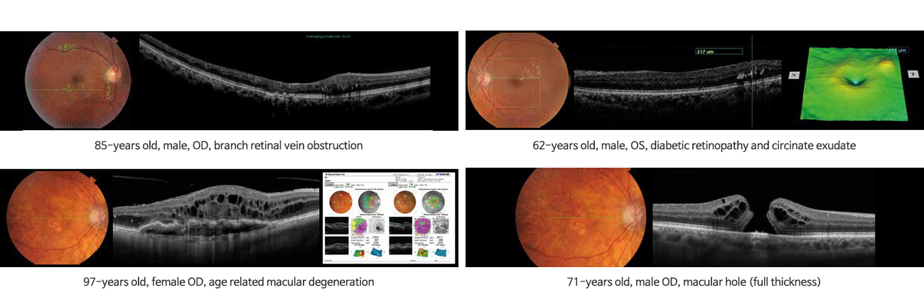
조직의 횡단면적 정보를 제공해 주어 망막 질환 및 조기 녹내장 진단에 유용한 첨단 진단 장비입니다.

망막 & 맥락막 진단
MACULA
-
- 5 Line cross scan
This scans with 5 line scan horizontally and vertically in an instant. This is useful for screening and for follow-up as it does not miss the target position by quick scanning.
-
- 3D Macula analysis
Horizontal box scan in macula area. 3D imaging is useful to understand the whole and precise form of the fovea area. Thickness map and normative database for retina thickness is available.
-
- Line scan
This enables high resolution B-scan with a maximum of 50 slices' overlapping.
-
- Radial scan
The radial scan is a fast solution to create an overview, with high resolution scans.
-
- True color fundus photography
The 3D OCT-1 Maestro has an integrated full color fundus camera. With one finger touch you can acquire simultaneously a posterior OCT image and a true color fundus image. This real fundus photo helps you quickly to locate the exact position of the OCT-scan and gives you additional information for diagnosis.
-
- Peripheral fundus photography
The 9-point fixation target in the 3D OCT-1 Maestro allows the operator to make 9 different color fundus photos and compose them into one total overview of the fundus. With optional software, a panoramic or mosaic overview can be created.
High quality/high resolution OCT and color fundus photography
Superb OCT technology
550.000 A-scans per second - More details in less time
A scanning speed of 50.000 A-scans/sec allows for faster tomography acquisition and produces clear cross-sectional retinal images. A clear, High-Definition, B-scan image is acquired with a high speed of 50.000 A-scan/sec by the simplest operation ever.Wide field OCT scan
With the 3D OCT-1 Maestro you can produce the perfect overview capture in a single image. The 12x9mm wide field OCT scan for the optic nerve & macula is perfect for fast screening. (image of 12x9mm)시신경 & 녹내장 진단
GLAUCOMA
-
- 3D Wide scan (12x9mm)
This allows to screen from the fovea to the optic nerve by single scanning. Thickness maps of RNFL, GCC and retina are available.
-
- 3D Disc analysis
Disc topography which combines fundus photography, various peripapillary parameters and RNFL thickness is available. The normative database for RNFL is also incorporated.
-
- 3D Macula (V) glaucoma analysis
Vertical box scan in macula area. GCC analysis is available and normative database for RNFL, GCC and retina thickness is incorporated.
-
- Color fundus photography / peripheral fundus photography
Non mydriatic color fundus photography is possible. The report template is ready for color fundus photography. Peripheral fundus photography is also available.
전안부 진단
ANTERIOR
-
- Anterior radial scan*
This allows to check the central cornea condition in 12 radial scan. Corneal curvature map and corneal thickness map is also available.
-
- Anterior line scan*
This allows to observe the Angle area.
*Anterior scanning is optional with anterior segment attachment (HA-2).


- 광주스마일안과의원
- 대표 : 조용윤
- 사업자등록번호 : 552-92-00700
- 광주광역시 서구 상무중앙로 58 (상무지구 BYC사거리 타임스타워빌딩 4층)
- TEL 062-381-3900
- FAX 062-381-9800